
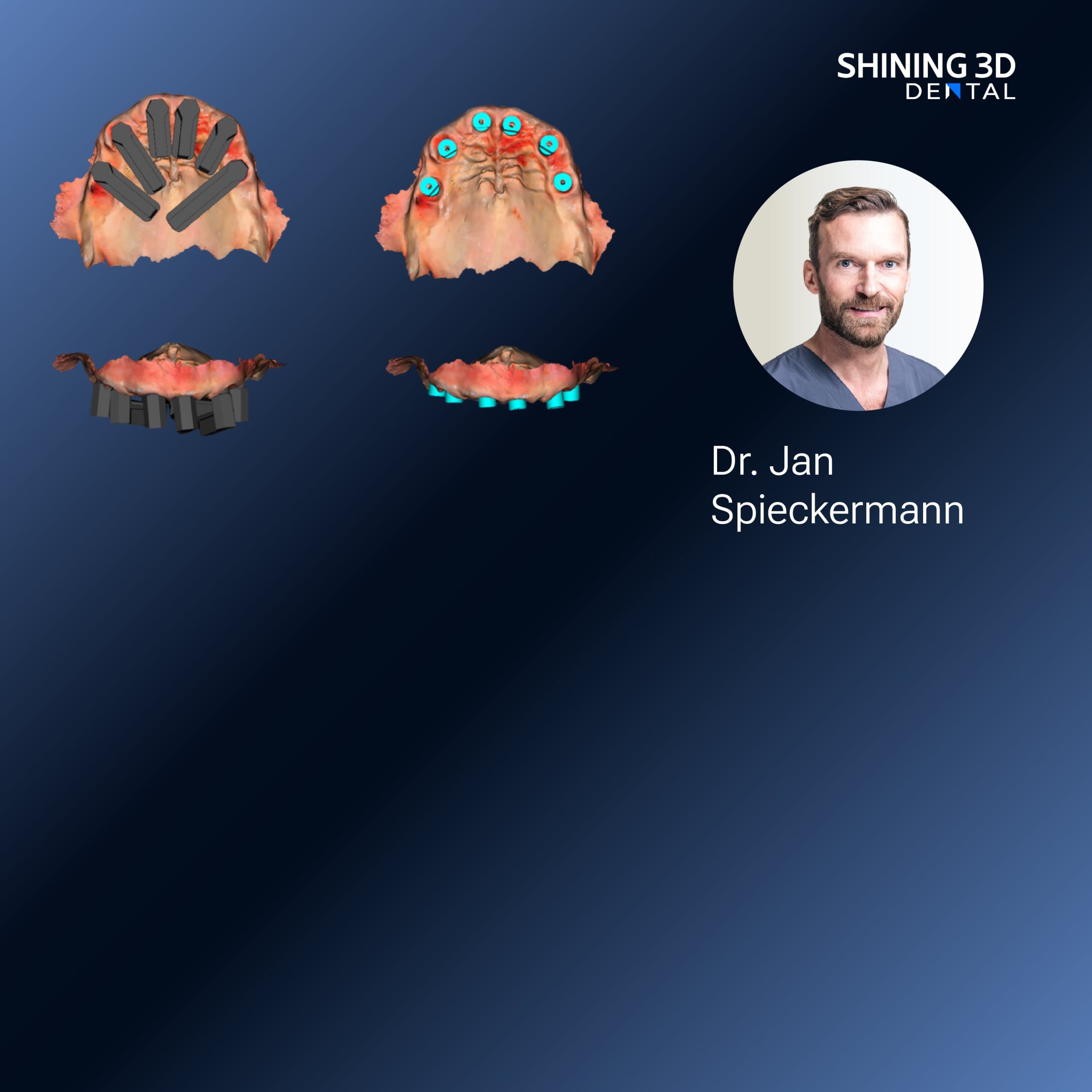
When scanin’ goes wrong — the hard knocks first
Folks come in thinkin’ a shiny camera’ll do all the work, but it don’t — bad light, bad stance, and sloppy files’ll wreck a session faster’n a wet barn roof on a windy night. I’ve watched setups choke on glare, lose texture, and spit out meshes that won’t mate with lab software. If you use a Photogrammetry 3D scanner, you still got to mind the basics: steady, even light; predictable head position; and file habits that don’t bury the good scans. That’s the problem we gotta fix first.

Light: stop fightin’ the sun and tame yer highlights
Hard shadows and hot spots confuse algorithms. Aim for soft, even illumination from two diffused sources at 45-degree angles to the face; no single overhead lamp, no bright window behind the subject. Use neutral color temperature (around 5000–5500K) so skin tones and textures stay honest. If teeth flash like tin, slap on a polarizing filter or lower specular highlights in capture settings. Check histograms — if highlights clip, the texture’s gone and no software’ll invent it back.
Positioning: get the head where the software expects it
Ask the person to sit tall and breathe steady. Use a plain background, remove glasses and dangly jewelry, pin hair back. Keep the camera distance consistent — too close, you miss context; too far, you lose fine texture. Move the scanner (or camera) in smooth arcs: corners of the eyes, bridge of the nose, cheek sweeps, chin undercuts. Small rotations matter more than frantic circling. If you need a reality check, remember what vendors showed at the IDS (International Dental Show) in Cologne — the demos that worked were the ones where the operator held steady and kept angles predictable while testing intraoral photogrammetry / IPG.
File management: name, save, and trim like you mean it
Don’t let meshes rot in Downloads. Give files sensible names (patientID_date_site), keep an original raw capture, and export trims only after you verify scale and normals. Export formats depend on use: PLY for color+geometry, OBJ when you want wide compatibility, STL for pure geometry. Before sending to the lab, run a quick pass: check for flipped normals, fill tiny holes under 1–2 mm only, decimate heavily only when file size blocks transfer but keep a full-res master. Version everything so you can go back when a problem shows up downstream.
Common pitfalls and the quick fixes that actually work
Movement blur — slow your workflow, calm the subject, use shorter capture bursts. Shiny teeth and wet mucosa — dry the area, use a matte spray if acceptable, or capture extra reference frames and mask specular areas later. Hair and scarves cause stray geometry; remove them or clip them in the first pass. Mismatched color between frames — lock white balance and keep the same light positions. If automatic alignment fails, add a few small, removable markers to give the algorithm anchor points.

Alternatives, when to switch tools, and what to expect
If you need intraoral detail at tooth-level accuracy, dedicated intraoral photogrammetry or structured-light intraoral scanners outperform face-focused photogrammetry for cramped oral cavities. For full-face texture with lower hardware cost, multi-camera photogrammetry rigs work but demand stricter lighting control. When speed is king and repeatability matters, go with devices and workflows that force consistent capture steps rather than leaving it all to operator luck.
Wrap-up: steady habits beat fancy kit every time
Fix the three big leaks — light, positioning, and file discipline — and your captures stop givin’ you headaches. These fixes are what separates a one-off lucky scan from a dependable workflow, and they’re the sort of practical details baked into respected systems from SHINING 3D DENTAL, so your scans actually move clean from chair to lab without needless back-and-forth.